| Home | Manuals | Software | References | Education | Research/Validation | Support |
|
|
|||||||||||||
|
|||||||||||||
| Cryomicrotome | |
|
A new system that directly determines the spatial location of every
fluorescent microsphere within organs of small laboratory animals has
been designed, reported and validated (Kelly, et al). This newly
developed Imaging CryoMicrotome allows mathematical dissection of
organs, and introduce new methods for characterizing and visualizing
spatial distributions of blood flow in small laboratory animals. |
|
|
The Imaging CryoMicrotome (Barlow Scientific, Inc. Olympia WA) consists
of a CCD video camera, a metal halide lamp, excitation filter-changer
wheel, emission filter-changer wheel, and a cryostatic-microtome (figure 1).
Fluorescence images are acquired with the digital camera.
Two motorized filter wheels containing excitation and emission filters
are mounted in front of the light source and camera, respectively. A
custom designed microtome serial sections through frozen organs.
Computer control of the microtome motor, filter wheels, and image
capture and display is accomplished through a virtual instrument
written in LabVIEW 5.1 (National Instruments Inc.). |
|
|
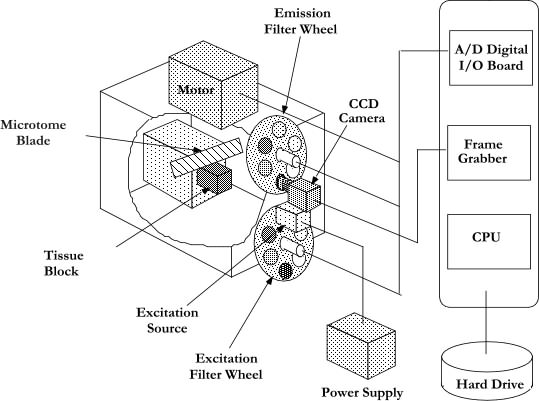
Figure 1. Schematic Diagram of Imaging CryoMicrotome. The Imaging CryoMicrotome (Barlow Scientific, Inc. Olympia WA) is a device that determines the spatial distribution of fluorescent microspheres at the microscopic level. The system collects microcirculation data from organs containing up to four different colors of fluorescent microspheres. The instrument consists of a CCD video camera, a metal halide lamp, excitation filter-changer wheel, emission filter-changer wheel, and a cryostatic-microtome. Fluorescence images are acquired with the digital camera. Two motorized filter wheels containing excitation and emission filters are mounted in front of the light source and camera, respectively. A custom designed microtome serial sections through frozen organs. Computer control of the microtome motor, filter wheels, and image capture and display is accomplished through a virtual instrument written in LabVIEW 5.1 (National Instruments Inc.). |
|
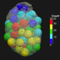
The CryoMicrotome serially sections through the frozen organ at a selected slice thickness. Digital images of the tissue surface (en face) are acquired with appropriate excitation and emission filters to isolate each of four different fluorescent colors (see heart and lung composite figures below). Each image is processed so that X and Y locations of each microsphere are determined. The spatial resolution of the system depends on the size of the organ being processed. Images of organ cross-sections produce a three-dimensional binary map defining the spatial location of organ parenchyma. This map determines the organ space locations to be sampled and the three-dimensional space for the statistical sampling. Acquired images are analyzed with software written in LabVIEW which writes the spatial coordinates and cluster size of each microsphere to a text file. Data reduction is automated through a final analysis program written in C, which removes camera artifacts and prevents microspheres being counted twice if they exist in more than one consecutive slice. Selected References:
|
|
|
Movies |
Graphics |
|||
|
|
||||
|
Rabbit Heart (6.4M) |

|
Heart Composite ( 60K) |

|
|
|
Rat Lung (7.5M) |
|
Lung Composite ( 58K) |

|
|
|
Outline Movie (4.7M) |
|
Lung and Heart Slices (117K) |

|
|
|
Mouse Heart Movies |
|
|
|
|